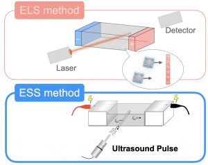
Figure 1: In contrast to electrophoretic light scattering, which uses a laser to measure particle motion, the electrophoretic dynamic ultrasound scattering (ESS) method uses ultrasound pulses.
When the surface of a particle is positively or negatively charged, applying an electric field to the particle suspension causes the particles to move to electrodes of opposite polarity, depending on the charge state of the surface. This phenomenon is well known as electrophoresis, and the measurement of electrophoretic mobility reveals the potential (charge state) of the particle surface, called zeta potential. The electrophoretic light scattering (ELS) method is often used for this measurement. Measurement of samples with high concentration is difficult with ELS (although there is a method to make the beam slightly submerged, but it is limited in size and concentration, and the accuracy is more or less insufficient), but with ultrasonic waves, optical turbidity and multiple scattering of light do not interfere with the measurement. For example, the electric sonic amplitude (ESA) method, which reads changes in ultrasonic amplitude caused by an electric field, and the colloid vibration current (CVI) method, which reads changes in electric current generated by ultrasound, have been reported in previous studies as ultrasound-based techniques. On the other hand, these methods have left some issues such as the fact that the results are dependent on the particle size, i.e., the particle size must be measured correctly in advance, and a reference is required. In addition, because the frequency of ultrasound is in the megahertz band, the electrophoretic mobility obtained was limited to information called dynamic electrophoretic mobility, which is restricted to high frequencies.
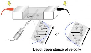
Figure 2: Near the cell wall, a flow in the opposite direction to that of the particles occurs, called electroosmotic flow, and the ESS method uses phases to acquire data at different sample positions in a single measurement without any repeated scanning measurements. It can also identify the direction of particle motion, as well as the direction of motion that is positively or negatively inverted due to osmotic flow.
We have developed an electrophoretic dynamic ultrasound scattering (ESS) method that “directly reads” the motion of particles in a liquid when an electric field is applied, and have analyzed the zeta potential of particles in very high concentration suspensions of particles (~50 wt%) . In this paper, we discuss the application of ESS to micron particles (2.5 μm), which are difficult to measure by the ELS method. This is similar to the difficulty of measurement when aggregates are included. The ESS method has the unique advantage of being able to detect not only the scattering amplitude of ultrasonic waves but also their “phase”. This allows us to lock-in and extract only the information on the electrophoretic motion from the two motions, “sedimentation” and “electrophoresis”. In addition, by making full use of the phase, positional information in the sample can be easily extracted in a single measurement, allowing visualization of the aforementioned electroosmotic flow profile. We have studied negatively charged silica as well as mullite particles with polarity inversion between acidic and basic conditions as an example. As a result, we were able to visualize not only the quantitative evaluation of electrophoretic mobility including the positive and negative polarity of the particles, but also the polarity (direction of motion) inside the sample due to electro-osmotic flow.
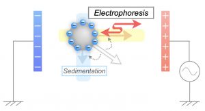
Figure 3 Sedimentation effects are often problematic for micron-sized particles and often-occurring aggregates. The ESS method uses a unique technique called the lock-in method to discriminate between sedimentation motion and electrophoresis, allowing only the electrophoretic information to be extracted.